Author: Benoit Dupont
All patients know that X-ray is a black and white image, right? And what if it would be possible to extract color information out of the image? Not for the sake of the cosmetic aspect of the picture, of course, but what if it had some diagnosis value, for the clinician? Well, it turns out it does.
• Caeleste Background
Caeleste is a begian CMOS image sensor expert design house (www.caeleste.be ). we introduced a new way to realize X-ray imagers based on energy separation (also called “color X-ray”). We are working closely with radiologists and researchers on this topic to see if it really makes sense to have such color detectors. We published early results on the value of energy differentiated color X-ray for mammography in ref [ ] and recently, researchers have proven in a publication at ESR2011 in Vienna (ref [ ]) that color X-ray applied to mammography would lead to a better diagnosis of tumor stages and Multifocality of lesion.
• Is color X-ray imaging interesting or just a hype?
We are not doctors but physicists and working with photons. When it comes to X-ray photons, their absorption by a material depends on its atomic weight. Color X-ray imaging is a technique where spectral information (“color”) is generated based on X-photon absorption versus energy differentiation. The color component in these images contains extra information: the chemical nature of the absorbing tissue.
How does this work? Next graph is the atomic composition of various body tissues and fluids:
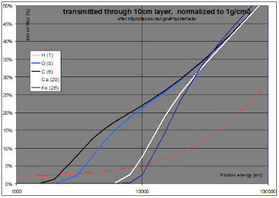
The absorption of X-rays by any material depends on the atomic weight of its composing elements. This absorption itself depends also on the wavelength, hence photon energy, of the X-ray. Acquiring an image for different wavelengths will thus reveal the atomic composition of the material. Color X-ray may add significant diagnostic value to screening or diagnostic mammography. This is in the first place due to the clear separation of C-rich fat from O-rich (vascular, glandular and collagenous interstitial) tissues, and by the enhanced visibility of Ca-rich calcifications and tumors in a C- and O-rich background. The following graph shows the comparative spectral absorption of X-rays for key body elements (H, O, C, Ca, Fe):
All medical X-ray can benefit from true color X-rays, ranging from general radiography, mammography to CT.
Radiologists know that color images can be made on classical digital X-ray machines. That technique is called dual energy X-ray imaging. One takes successively two images at two different X-ray energies. The results are pixel-by-pixel processed to yield color X-ray images.
Dual energy x-ray imaging suffers from two drawbacks: the additional x-ray image results in an elevated patient dose and the slower temporal resolution of the system may cause image registration problems due patient/organ movement.
Photon counting detectors do not suffer from that dose increase. Photon counting detectors count and sort in each pixel the photons based on their energy. It inherently does not need any more photons that those already there for the B&W absorption X-ray image.
Figure above: B&W X-ray absorption and color X-ray images from the same mammary resection specimen (from [reference 1]). In this color image, hue codes the oxygen/carbon ratio (here blue=carbon-rich, red=oxygen-rich)
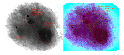
Figure above: B&W X-ray absorption and color X-ray image of cancerous tissue specimen. Color image was image processed to enhance edges. Central: malignant tumor with central calcifications. Extensions: fibrous tissue seem to contain foci of incipient cancer; extensions at 10 and 7 o’clock reach the resection margin.
• Conclusion
Such low dose color imaging is only possible with CMOS detectors, that will in the end replace black and white TFT plates. Such sensors are under development at Caeleste to be integrated in next generation X-ray imaging equipment. Caeleste’s concepts and patents bring such sensors to a manufacturing cost close to TFT plates.
Caeleste will also continue his work with academic world, researcher and diagnosticians to bring the best possible diagnosis solutions.
• About Caeleste
Caeleste is a Belgium based start-up founded in 2006 designing and prototyping high-end image sensors, in particular photon counting X-ray image sensors. Caeleste’s founders are experts in the image sensor and radiation detector domains, totalizing more than 150 academic publications and 50 international patents. Based in Europe, Caeleste operates worldwide.
www.caeleste.be
• About the author
Benoit Dupont is co-founder and design-lead at Caeleste. He received his PhD from Paris Sud XI University in 2008 and his engineering degree in Microelectronic from ISIM, Université Montpellier II in 2002. He is also invited teacher at Paris XIII University.
Benoit.dupont@caeleste.be
References:
[1] B. Dierickx, N. Buls, C. Bourgain, C. Breucq, J. Demey, B. Dupont, A. Defernez, “On the diagnostic value of multi-energy X-ray imaging for Mammography”, European Optical Society symposium, 16-18 June 2009, Munchen
[2] I. Willekens, B. Dierickx, N. Buls, C. Breucq, A. Schiettecatte , J. de Mey and C. Bourgain “Superiority of multi-energy color X-ray for breast specimen radiography.” European Society for Radiography, Vienna, March 2011


the reference publications did not go through the back-office of the website, it seems. I paste them here in the comments:
ReplyDelete[1] B. Dierickx, N. Buls, C. Bourgain, C. Breucq, J. Demey, B. Dupont, A. Defernez, “On the diagnostic value of multi-energy X-ray imaging for Mammography”, European Optical Society symposium, 16-18 June 2009, Munchen
[2] I. Willekens, B. Dierickx, N. Buls, C. Breucq, A. Schiettecatte , J. de Mey and C. Bourgain “Superiority of multi-energy color X-ray for breast specimen radiography.” European Society for Radiography, Vienna, March 2011